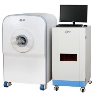
How A 1.0T Small Animal NMR Brings The Benefits for Preclinical Research?

Magnetic resonance imaging instrument (MRI), is an important equipment in most health centers. It is used to photo the tumor or disease inside the patient's body. MRI has its benefit of non-invasive, zero radiation, image clarity, fast imaging, and other advantages. Compares to X-ray Scanner or Computed Tomography Scanner (CT Scanner, it is also a kind of X-ray machine), MRI Scanner is a very safe instrument. So far, there is nothing hazard to human body was found.
For most of the life science research, biotechnology, pharmaceutical new medicine development, clinical trial applications, MRI is a very ideal device, it can carry out in pre-clinical animal experiments. However, this kind of human body MRI, typically costs million dollars, not to mention the maintenance and consumables, such as liquid helium supplies. For the general laboratory in the university, MRI is an luxury expensive equipment, and it's difficult to equip.
At present, there have been some small form factor MRI for pre-clinical research in the market. This type of MRI is typically from 7T (Tesla, magnetic field strength unit), 3T, 1T, even less than 1T MRI. Among these small form factor MRI, those under 3T low field MRI, using high-precision permanent magnets, helium free design. It reduced the maintenance cost close to zero.
NM-G1 1T Small Animal MRI Scanner, from NIUMAG, provides a 40mm diameter probe, which is sufficient for small animals such as mice, and it can take the section profile photographs of the animals body. One can observe the body dynamically, and take persistent imaging, without any hurt to the animals being scanned. Because of the lack of operating costs, NM-G1 1T Small Animal MRI Scanner is possible to observe the small animals with a low-cost, non-invasive manner for a long time. It is used to understand the effectiveness or harm of drugs, micro poisons or pests for a period of time.
Summarized the benefits of NM-G1 1T Small Animal MRI Scanner for preclinical studies:
- Small animal section profile imaging, for the observation of specific visceral volume size. For example, the size of the kidney.
- Tumor size, continuous tracking drug treatment of the tumor effect
- Fat distribution. Obesity, nutrition and other related animal research
- Pharmacological. The role of nano-drug carrier in vivo, and metabolic rate assessment, targeting drug carrier targeting to determine
- Contrast agent imaging: contrast agent in vitro imaging
- All other studies that requires prolonged observation of changes in small animals.


